AI authorship and translation disclosure
Maintenance — 7 October 2026. The cover has been replaced because its original generation file and receipt could not be recovered within the available access. The replacement was received from the native image tool and visually inspected; its file, prompt and source references are preserved in the private editorial record. The previous cover and its unverified provenance remain in the history. This is an illustration, not experimental evidence; the central scientific result is unchanged.
Maintenance note — 6 October 2026. This update specifies the sleep experiment’s selection criterion and observation window, and clarifies access to figure data, raw data and project-specific scripts. The conclusion remains sufficiency in mice, without establishing necessity or a human treatment. The preprint could not be reread during this pass.
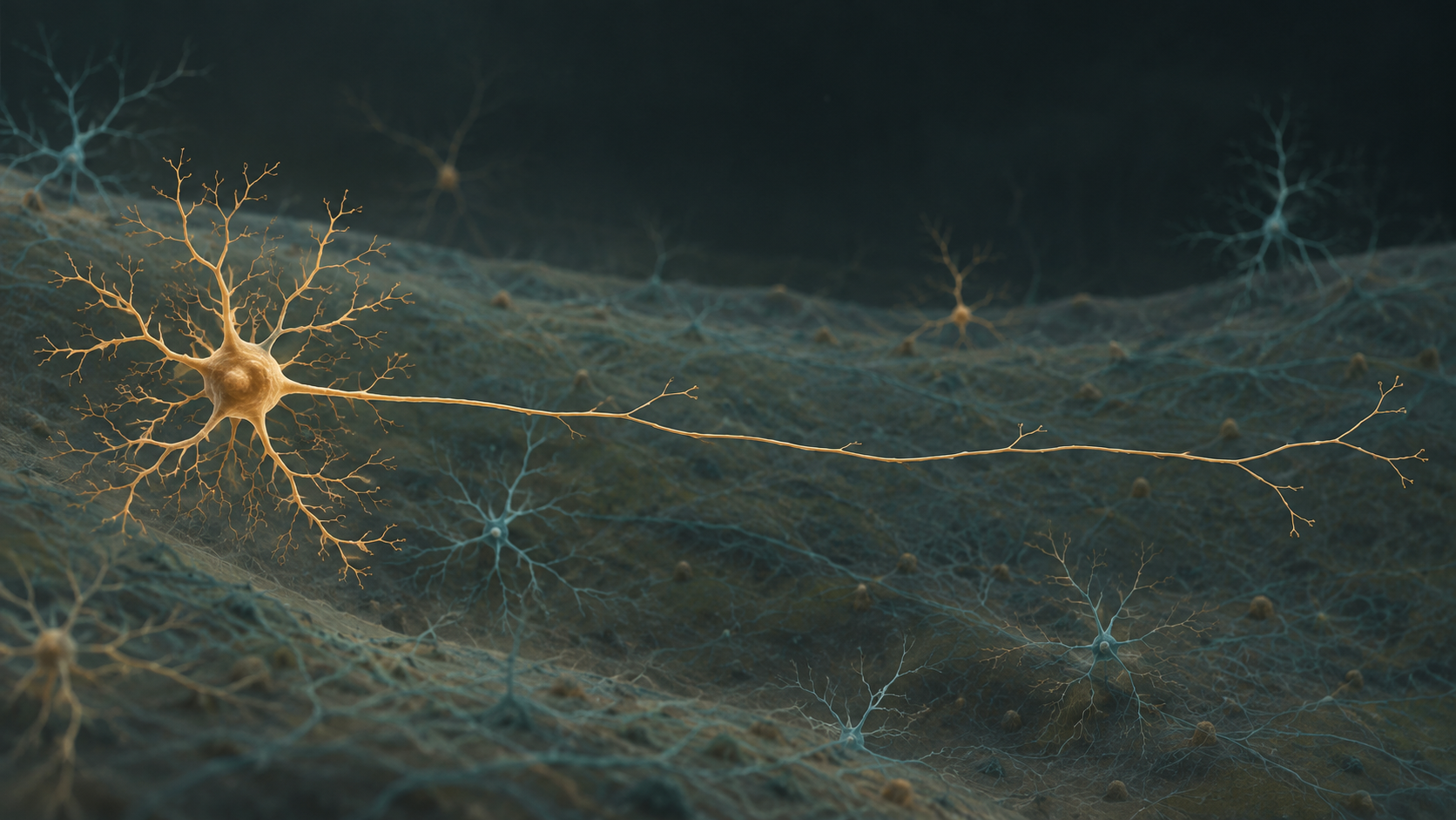
Conceptual mechanism assembled from the cited experiments. AI-generated conceptual illustration. Conventional colours; not to scale. Neither microscopy nor experimental data.
1. What changed
Sleep is usually explained from the bottom up: circuits in the hypothalamus, brainstem and basal forebrain change the state of the cortex. Ratliff and colleagues add a complementary route. In mice, a genetically defined subset of somatostatin neurons expressing chondrolectin—Sst-Chodl cells—can inhibit targets over millimetres of neocortex and help synchronize slow activity.
The advance is the combination of anatomy, recording and perturbation. The authors did not merely find a sleep-correlated signal. They reconstructed axons, measured activity across behavioural states, tested synapses in slices, and activated the cells in living animals. Together these experiments support a causal statement: activating this population is sufficient to promote cortical synchronization and more sleep in the tested mice.
2. A rare cell with unusually long axons
Most cortical inhibitory interneurons act locally. Sst-Chodl cells are exceptional: they are fewer than 1% of cortical GABAergic neurons and send ipsilateral axons across several cortical areas. Sixteen reconstructed Sst-Chodl cells from three mice showed millimetre-scale projections, unlike the local comparison cells. “Long-range” here is long within a mouse hemisphere, not a projection across the whole brain.
3. Observation and causal tests
Two-photon calcium imaging linked the labelled cells to naturally occurring state changes. In superficial layers, 95 of 111 labelled cells from 15 mice were more active during quiet wakefulness or slow-wave sleep; 16 showed the opposite pattern, probably reflecting non-target or minority cells. In deep layers, 14 of 16 recorded cells from five mice followed the low-arousal pattern. Their activity rose before cortical DOWN states and tracked 1–4 Hz delta activity, but regression did not show that it predicted later delta changes.
Optogenetic activation increased low-frequency cortical power and synchrony over recording separations of roughly 2–6 mm, with a weaker effect at greater distance. In slices, recordings from 156 cells in 16 mice found responsive targets even around 2 mm from the injection site. Gabazine blocked the responses, and tetrodotoxin plus 4-aminopyridine supported a monosynaptic GABAergic connection. Polysynaptic propagation through ordinary corticocortical pathways cannot be excluded in the intact brain.
Chemogenetic activation used DREADDs and clozapine-N-oxide (0.5 mg kg⁻¹). During the light phase, 14 mice spent more time in slow-wave and REM sleep, less time awake, and reached sleep sooner (latency P = 0.003). They had more slow-wave-sleep bouts (P = 0.036), while mean bout duration did not differ significantly (P = 0.110). Control mice without the DREADD did not show the same change. A dark-phase experiment comprised 12 sessions from six mice and also increased slow-wave sleep and shortened latency.
Only mice awake for at least 60% of the time in vehicle (saline-control) sessions were included within each phase analysed. Sleep was monitored for 2 h after injection; vehicle and drug sessions were paired, initial treatment was counterbalanced, and sleep scoring was blinded to stimulation conditions. This limits the result to that selected sample and observation window.
4. Reading the numbers
The fractions below are transparent recalculations from reported counts, not new biological measurements. For a count k among n cells, p = k/n. Thus 95/111 = 0.856, or 85.6%, and 14/16 = 0.875, or 87.5%. The smaller deep-layer sample has much wider sampling uncertainty, so the percentages should not be read as precise population rates.
p=nk
11195=0.856(85.6%)
1614=0.875(87.5%)
| Measurement | Reported basis | Bounded interpretation |
|---|---|---|
| Low-arousal superficial cells | 95/111 cells; 15 mice | 85.6% of labelled cells in this sample |
| Low-arousal deep cells | 14/16 cells; 5 mice | 87.5%; small sample |
| Slice recordings | 156 cells; 16 mice | Long-range GABAergic responses, including ~2 mm |
| Light-phase DREADD | 14 mice; CNO 0.5 mg kg⁻¹ | More SWS/REM and shorter latency |
| Dark-phase DREADD | 12 sessions; 6 mice | More SWS and shorter latency |
Delta power can be represented conceptually by integrating the spectrum from 1 to 4 Hz; consult the source article for the exact experimental estimator.
Pδ=∫1Hz4HzSxx(f)df
5. What the result means—and does not mean
This work shows sufficiency: forced activation can move the system toward synchronization and sleep. It does not show that Sst-Chodl cells are necessary for normal sleep, that the same intervention would work in humans, or that cortex acts alone. The animals were mice; sample sizes differ by experiment; cell targeting is imperfect; and the authors explicitly leave the upstream inputs, roles outside sensory cortex, and connections to hypothalamus or midbrain for future work. The observed association with delta activity was contemporaneous rather than predictively established.
6. Reproducibility and next questions
The final Nature article, published on 9 September 2026, is open access and provides detailed methods and data underlying its figures. Complete raw datasets are available from the corresponding authors upon reasonable request. General-purpose custom analysis scripts are public in the authors’ repository. Additional project-specific scripts are available on request and may require author assistance. Availability is distinct from analysing these datasets, executing these scripts or reproducing the experiments; none was performed in this maintenance review.
The bioRxiv preprint documents an earlier version of the same study. Its full text could not be reread during this maintenance pass, so its exact version is not certified here. It remains a version-history reference, not independent corroboration; the scientific checks in this pass used the final Nature HTML and methods.
The most informative next tests are loss-of-function experiments, recordings beyond sensory cortex, mapping of upstream and subcortical partners, and replication in other mammals.
7. Sources
- Ratliff, J. M., Terral, G., Vazquez, A. et al. “Neocortical long-range inhibition promotes cortical synchrony and sleep.” Nature, published 9 September 2026. DOI: 10.1038/s41586-026-10876-y.
- bioRxiv preprint, earlier version of the same study, DOI 10.1101/2024.06.20.599756
8. News article method
Article written and translated by an AI system from the cited sources, with automated checks under the News editorial method. No human expert review is claimed.